Microscopes have been essential tools in scientific research for hundreds of years. They allow scientists to study and analyse objects at a microscopic level, leading to numerous significant discoveries in a variety of scientific fields, including biology, medicine, and materials science. In this article, we will take a deep dive into the fascinating history of microscopes, exploring their evolution over time and the impact they have had on scientific research.
The Origin
The origins of microscopes can be traced back to the 16th century, when eyeglass makers in the Netherlands began experimenting with lenses to create a device capable of magnifying objects. The first microscopes were rudimentary handheld devices that utilised a single convex lens to magnify the object being observed. These early microscopes were known as “simple microscopes.”
The first person to document the use of a microscope was Italian scientist Francesco Stelluti in 1625. Stelluti used a simple microscope to study the anatomy of a bee, observing its compound eyes and wings in great detail. However, it was Dutch scientist Antonie van Leeuwenhoek who is credited as the first person to develop a powerful and effective microscope.
Van Leeuwenhoek was an amateur scientist who made his microscopes by grinding lenses. His microscopes were small, with a single lens mounted in a brass holder. Despite their simple design, Van Leeuwenhoek’s microscopes were able to magnify objects up to 275 times their original size, far surpassing the capabilities of any other microscope at the time.
Van Leeuwenhoek used his revolutionary microscope to study a wide range of objects, from living organisms such as bacteria and protozoa to non-living materials such as crystals and minerals. His observations led to many important discoveries, including bacteria and red blood cells.
Evolution of Microscope Designs
Over time, the design of microscopes continued to evolve. In the 17th and 18th centuries, compound microscopes, which used two or more lenses to magnify objects, became more popular. These microscopes were often mounted on a tripod or stand and had an adjustable stage for holding specimens.
One of the most significant advancements came in the 19th century with the invention of the achromatic lens. Before this, microscope designs suffered from colour fringing and distortion, which limited the clarity of images produced. However, the achromatic lens was able to correct these issues, resulting in much clearer and sharper images.
Another significant development in the history of microscopes came in the 20th century with the invention of the electron microscope. Unlike traditional light microscopes, which use visible light to magnify objects, electron microscopes use a beam of electrons to create an image. This allows for much higher levels of magnification and resolution, making it possible to study objects at the atomic and molecular levels.
Electron microscopes come in two main types: Transmission Electron Microscopes (TEMs) and Scanning Electron Microscopes (SEMs). TEMs use a beam of electrons that passes through a thin specimen, creating an image on a fluorescent screen or detector. SEMs, on the other hand, scan a beam of electrons across the surface of a specimen, creating a three-dimensional image of its surface.
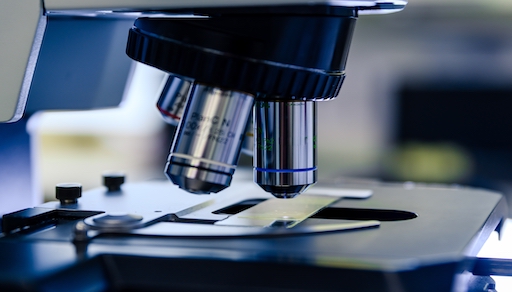
Today, microscopes are used in a wide range of scientific fields, including biology, medicine, materials science, and nanotechnology. There are many different types of microscopes available, each designed for a specific purpose. Some of the most common microscopes are optical microscopes, electron microscopes, scanning probe microscopes, and confocal microscopes.
Optical microscopes use visible light to magnify objects and are typically used to study specimens that are too large or opaque to be studied using electron microscopes. They are also commonly used in biological research to study cells and tissues. Confocal microscopes are a type of optical microscope that uses lasers to illuminate a specimen and create a three-dimensional image.
Scanning probe microscopes are a type of microscope that uses a probe to scan a surface and create an image. They are commonly used in materials science to study the surfaces of materials at the atomic and molecular levels. They are also used in nanotechnology research to manipulate and measure nanoscale structures.
In conclusion, the history of microscopes is a fascinating journey that has led to many important scientific discoveries. From the simple handheld devices of the 16th century to the sophisticated electron microscopes of today, microscopes have greatly expanded our understanding of the world around us. They continue to be an essential tool in scientific research and discovery, with countless discoveries yet to be made. At Techmate, we offer an extensive range of microscopes that can assist you in your experiments. Select a microscope from our catalogue or contact us for assistance.